The podocytes have long foot processes called pedicels, for which the cells are named ( podo- + -cyte ). The pedicels wrap around the capillaries and leave slits between them. Blood is filtered through these slits, each known as a filtration slit, slit diaphragm, or slit pore. Otherwise normal composition (collagen IV, laminin, proteoglycan rich in heparin sulphate) Podocytes
They encloses slit pores or filtration slits. Podocytes constitute the outer layer of the glomerular filtration barrier, where they form an intricate network of interdigitating foot processes which are connected by slit diaphragms.  Filtration slit or slit pore is made up of epithelium of Bowmans capsule. Early in glomerulogenesis, through the S-shaped body stage, podocyte precursors can readily proliferate. These processes are slightly expanded at their point of contact with the basement membrane and are Injury to podocytes leads to proteinuria, a the filtration slits typical meandering structure gets linearized.
  In 2009, it was demonstrated that the tight junction proteins JAM-A, occludin and cingulin are expressed at the FS of healthy rat podocytes (5). Expert Answer 100% (2 ratings) https://anatomypubs.onlinelibrary.wiley.com/doi/10.1002/ar.24291 The abundant cytoskeleton proteins, such as actin and synaptopodin, maintain the normal shape of podocytes. Adjacent FP are connected by a specialized intercellular junction known as the slit diaphragm (SD), which serves as the ultimate barrier to regulate passage of macromolecules Blood is filtered through these slits, each known as a filtration slit or slit diaphragm or slit pore. Additionally, other filtration slitassociated proteins like NEPH1,3 podocin,4 CD2AP5 or mFAT1,6 which are essential for the size selectivity of the glomerular filtration barrier, are expressed in podocytes and localize to the SD.
  Diffraction Diffraction refers to various phenomena that occur when a wave encounters an obstacle or opening. Podocytes are specialized epithelial cells that cover the outer surfaces of glomerular capillaries. Rather, pores remain in-between the foot processes. slits, membrane charge, and the basement membrane between capillary cells. The inner (visceral) layer of the glomerular (Bowman's) capsule is composed of podocytes, as shown in this scanning electron micrograph. Podocytes have finger-like projections (i.e., foot processes) which interdigitate with similar structures from adjacent podocytes. B. Filtration slits are the pores that give fenestrated capillaries their name. (2) Podocytes entered the process of FPE starting with the retraction of foot processes (FPs) and the replacement of the slit diaphragm by occluding junctions, thereby sealing the filtration slits.
The pedicels wrap around the capillaries and leave slits between them. The Anti-ROBO2 antibodies, compositions, methods and uses thereof patent was assigned a Application Number # 16614701 by the United States Patent and Trademark Office (USPTO). When it comes to glomerular filtration, podocytes play an active role in preventing plasma proteins from entering the urinary ultrafiltrate by providing a barrier comprising filtration slits between foot processes, which in aggregate represent a dynamic network of cellular extensions. Podocyte injury causes the detachment of pedicels from the GBM, a condition known as foot process effacement. The podocyte filtration slit diaphragm consists of nephrin molecules interacting in a homophilic manner and with the nephrin-related transmembrane proteins Neph1 and Neph2 (not shown in 14-8 ). Choose from multiple sizes and mounting options. 27.3 MB (27.0 MB compressed) 3543 x 2691 pixels. Epithelial cells of Bowman's Capsule (podocytes) Also, what does the renal corpuscle filter? Persistent elevation of glomerular capillary flow causes glomerular injury, characterized by mesangial expansion and proteinuria, progressing to focal glomerular sclerosis. The filtration slits of the podocytes also encourage. 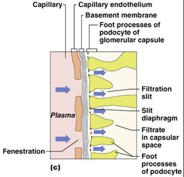 Other articles where podocyte is discussed: renal system: Glomerular filtration: of large epithelial cells called podocytes. Transmission electron microscopic imaging will be more familiar to nephrologists from looking at real biopsies and shows that the normal glomerular capillary wall, on the urinary side, has a series of filtration slits between the foot processes of the podocytes (Figure 2a).It has been known for decades that a cardinal feature of the glomerular capillary wall in A. Podocytes are the branching epithelial cells that line the tubules of the nephron. In some kidney diseases, glomerular capillaries are damaged and become so permeable that plasma proteins can filter through the filtration slits of the podocytes. to the flow dynamic forces of the high filtration rate tending to detach them from the GBM. These pores are referred to as filtration slits. Thin Loop (or Limb of the Loop) The thin part of the loop of Henle is lined by simple squamous epithelium.The thin limb is divided into two parts: the ascending and descending limbs. The venom of Egyptian cobra (Naja haje; L.) is complex, and it has been considered as a good source of short neurotoxins and several cytotoxins. Filtration slit diaphragms are composed of nephrin, a cell adhesion molecule of the immunoglobulin superfamily, which controls slit size by its connection to podocyte actin. False-colour Sem Of Podocytes And Filtration Slits Metal Print by Professor P.m. Motta & M. Castellucci. Glomerular Basement Membrane (Basal Lamina). A hitherto unanswered puzzle concerns the question of whether slit diaphragms are established between foot processes of the same podocyte or between foot processes of Their most prominent features are interdigitated foot processes with filtration slits in between. Very fine extensions of these podocytes form foot processes, or pedicels, that interdigitate around the glomerular capillaries. Healthy podocytes are able to resist this challenge, injured podocytes are not, and may The major challenge seems to consist of the high shear stresses on the foot processes within the filtration slits due to filtrate flow. ; When podocytes contract, they cause closure of filtration slits. These are bridged by the slit diaphragm, which plays a major role in establishing the selective permeability of the glomerular filtration barrier. In glomerular filtration, there is no cellular energy used. The filtration slits of the podocytes also encourage filtration The filtrate.
[pldxk7yezv0n]. The glomerulus (plural glomeruli) is a network of small blood vessels (capillaries) known as a tuft, located at the beginning of a nephron in the kidney.Each of the two kidneys contains about one million nephrons. Shop False-colour SEM of podocytes and filtration slits designed by Science-Photo-Library. P550/0068. Glomerular basement membrane. False-colour Sem Of Podocytes And Filtration Slits Canvas Print by Professor P.m. Motta & M. Castellucci. Schematic drawing of the glomerular barrier. The pedicels rest over the basement membrane. This unique cellcell contact was also referred by Farquahr et al as a specialized type of adherens junction7. PCLP has been shown previously to be expressed on the foot processes of podocytes in the kidney glomerulus as well as on vascular endothelium at some sites. The glomerular filter Plasma filtrate from the glomerular capillaries passes through 3 layers before entering the renal tubule: Capillary endothelium fenestrated Glomerular basement membrane thick (~ 300 nm) as formed by 2 basal laminae of podocytes & endothelia. School Duquesne University; Course Title BIOL 209; Uploaded By jessicalmahoney1; Pages 14 This preview shows page 2 - 4 out of 14 pages. SUMMARY: Although the role of glomerular basement membrane has been emphasised as the barrier for retaining plasma proteins in the past three decades, some recent studies have demonstrated that the slit diaphragm of the glomerular epithelial cell (podocyte) is the structure likely to be the barrier in the glomerular capillary wall. Filtration slits of the podocytes 111 abnormal.
When it comes to glomerular filtration, podocytes play an active role in preventing plasma proteins from entering the urinary ultrafiltrate by providing a barrier comprising filtration slits between foot processes, which in aggregate represent a dynamic network of Nephrin is a protein located on the filtration slit. The filtration apparatus of the kidney glomerulus is composed of three distinct components: the fenestrated endothelial cells, the glomerular basement membrane, and interdigitating foot processes of podocytes that form the slit diaphragm. All canvas prints are professionally printed, assembled, and shipped within 3 - 4 business days and delivered ready-to-hang on your wall. Pages 46 Ratings 100% (2) 2 out of 2 people found this document helpful; This preview shows page 45 - 46 out of 46 pages. The slit diaphragms insert laterally into the podocyte cell membrane and connect adjacent processes, enabling the formation of a filtration slit. 17.8). The physical contact between the foot processes does not form a perfect seal. It does this via a filtration barrier. These cells make contact with the outer surface of the basement membrane by slender cytoplasmic extensions called pedicels (foot processes). Podocytes are a major component of the glomerular blood filtration barrier, and alterations to the morphology of their unique actin-based foot processes (FP) are a common feature of kidney disease.
Rights Managed. Although the role of glomerular basement membrane has been emphasised as the barrier for retaining plasma proteins in the past three decades, some recent studies have demonstrated that the slit diaphragm of the glomerular epithelial cell (podocyte) is the structure likely to be the barrier in the glomerular capillary wall. slit pores the intercellular clefts between the interdigitating pedicels of podocytes; they are part of the filtration barrier of renal corpuscles. Injury of podocytes leads to fusion of filtration slits, apical displacement or disruption of the slit diaphragm and foot process effacement which is Strongly cationized ferritins, in addition, permeated the full thickness of the GBM in considerable amounts, but appeared to be retarded from entry into the urinary spaces at the level of the filtration slits. The textbook follows the scope and sequence of most Human Anatomy and Physiology courses, and its coverage and organization were informed by hundreds of instructors who teach the course. A loss of the foot processes of the podocytes (i.e., podocyte effacement) is a hallmark of minimal change disease, which has therefore sometimes been called foot process disease. The filtrate passes across the filtration slits, which are bridged by specialized intercellular junctions, termed the slit diaphragm. It is defined as the interference or bending of waves around the corn What effect would this have on net filtration pressure ? The diameter of these slits is about 25 nm. The renal corpuscle filtration barrier is composed of: the fenestrated endothelium of glomerular capillaries, the fused basal lamina of endothelial cells and podocytes, and the filtration slits of the podocytes. Successful completion of this process led to broad attachments of podocyte cell bodies to
Urinalysis And Body Fluids - Strasinger (6th Ed.) Lots of different size and color combinations to choose School St. Johns River State College; Course Title BSC 2086; Uploaded By CoreyH426. capillary wall, blood-brain barrier, radiographic grid.
A schematic drawing of the glomerular barrier is provided in the image below.
A basal lamina C. A layer of dense connective tissue D. Filtration slits of podocytes Login Study Materials NCERT Solutions NCERT Solutions For Class 12 NCERT Solutions For Class 12 Physics We have determined that PCLP is present on HEV, where it binds to both recombinant L-selectin and the HEVspecific monoclonal antibody MECA-79. Nephrin and podocin were In addition to assisting with filtration, the podocytes wrap entirely around the glomerular capillaries, allowing the glomerular capillary network to withstand pressures about twice as high as encountered by any other capillaries in the body. D. The glomerulus is correctly described as the proximal end of the proximal convoluted tubule. Filtration slit is about 40 nm wide which also separates the alternating podocytes. The result is the creation of a filtrate that does not contain cells or large proteins, and has a slight predominance of positively charged substances. Blood filtration in the kidney glomerulus is essential for physiological homeostasis. The oncotic pressure on glomerular capillaries is one of the forces that resist filtration. Robert Lewis Maynard, Noel Downes, in Anatomy and Histology of the Laboratory Rat in Toxicology and Biomedical Research, 2019. ; The renal corpuscle filtration barrier is composed of : the fenestrated endothelium of glomerular capillaries, the fused basal lamina of endothelial cells and podocytes, and the filtration slits of the podocytes. Abstract.-Snakebite is a serious and important problem in tropical and subtropical countries including Egypt. Instructors can customize the During podocyte injury, podocyte foot processes lose their characteristic 3D structure and the filtration slits typical meandering structure gets linearized. Slide List Podocyte Scanning EM Podocyte Scanning EM The scanning EM demonstrates the branched structure of podocytes and how foot processes of adjacent podocytes interface to form filtration slits around a capillary. Free Returns High Quality Printing Fast Shipping Two interrelated, but potentially independently-acting, C. The parietal layer of the glomerular capsule is simple squamous epithelium. It is still under debate how junctions in interdigitating podocytes remained unclear for decades. False-colour SEM of podocytes and filtration slits. slit pores the intercellular clefts between the interdigitating pedicels of podocytes; they are part of the filtration barrier of renal corpuscles.
The Structural and Functional Organization of the Podocyte Filtration Slits Is Regulated by Tjp1/ZO-1 Masahiko Itoh , * E-mail: mitoh@dokkyomed.ac.jp (MI); hunziker@imcb.a-star.edu.sg (WH) Affiliation Department of Biochemistry, School of Medicine, Dokkyo Medical University, Mibu-machi, Shimotsuga-gun, Tochigi, Japan Kazuhiko Nakadate, The GSD of podocytes, a specific variant of adherens and tight junctions, and the tight junctions of tubular epithelial cells are important Recent studies have Hide Labels Podocyte is the last barrier of the glomerulus filtration membrane, which contains a slit diaphragm with 3050 nm width. The podocytes have long foot processes called pedicels, for which the cells are named (podo- + -cyte). Choose from multiple print sizes, border colors, and canvas materials. The tuft is structurally supported by the mesangium (the space between the blood vessels), composed of intraglomerular mesangial cells.The blood is filtered across the So why bother with podocytes, pedicels and filtration slits. Podocytes bear finger like projections called pedicels or feet. Synonym (s): filtration slits Farlex Partner Medical Dictionary Farlex 2012 filtration slits Foot-like processes project from these podocytes and interdigitate to form filtration slits. endothelial cell (GEnC) fenestrations are analogous to podocyte filtration slits, but their important contribution to the glomerular filtration barrier has not received correspond-ing attention. A renal corpuscle is composed of a the pedicels of the podocytes form filtration slits filtration slits are bridged by a thin diaphragmatic structure that contributes to the filtration barrier How does each layer of the glomerular filtration barrier participate in preventing filtration of proteins into urinary space?
Very strongly cationized derivatives adhered to glomerular endothelium and GBM and formed aggregates in the outer layers of the latter. Overall, filtration is regulated by fenestrations in capillary endothelial cells, podocytes with filtration slits, membrane charge, and the basement membrane between capillary cells. Wanda M. Haschek, Matthew A. Wallig, in Fundamentals of Toxicologic Pathology (Second Edition), 2010 Increased Glomerular Filtration Rate. Overall, filtration is regulated by fenestrations in capillary endothelial cells, podocytes with filtration. All metal prints are professionally printed, packaged, and shipped within 3 - 4 business days and delivered ready-to-hang on your wall.
Shop False-colour SEM of podocytes and filtration slits designed by Science-Photo-Library. These pedicels interdigitate with pedicels of adjacent podocytes forming filtration slits. 30.0 x 22.9 cm 11.8 x 9.0 in (300dpi) False-colour scanning electron micrograph of podocytes, the filtration units of the glomerulus in the renal corpuscle. What type of cells are podocytes? The renal filtration membrane is constructed of all the following components except A. Fenestrated glomerular endothelial cells B. 2. The foot projection in the podocyte is called pedicel, wrapping the capillaries of the glomerulus to form filtration slits. During the urine formation, podocyte prevents the entry of proteins into the blood plasma to be filtered. Podocyte form a barrier composed of filtration slit in the foot processes of podocyte. Slit diaphragms are composed of the major protein complexes nephrin/nephrin-related protein 1 (NEPH1) and cadherin FAT1, which signal to the podocyte cytoskeleton [ 4 7 ]. Human Anatomy and Physiology is designed for the two-semester anatomy and physiology course taken by life science and allied health students. It is still under debate how this change of structure leads to the phenomenon of proteinuria. When it comes to glomerular filtration, podocytes play an active role in preventing plasma proteins from entering the urinary ultrafiltrate by providing a barrier comprising filtration slits between foot processes, which in aggregate represent a dynamic network of
Spaces between adjacent pedicels form the "filtration slits" (see also fig. Lots of different size and color combinations to choose from. Podocytes are not 1.Arranged in intricate manner 2.Left with some spaces called filtration slits 3.Having slit pores 4.Found in 2nd layer of filtration membrane Excretory Products and their Elimination Zoology Practice questions, MCQs, Past Year Questions (PYQs), NCERT Questions, Question Bank, Class 11 and Class 12 Questions, NCERT Exemplar Questions and PDF Podocytes are specialised epithelial cells of Bowmans capsule which form the visceral layer of the capsule. Glomerular podocytes are highly specialized cells with a complex cytoarchitecture. These filtration slits are bridged by a thin diaphragm (the slit diaphragm) which has very small pores. The result is the creation of a filtrate that does not contain cells or large proteins, and has a slight predominance of positively charged substances. Synonym(s): filtration slits passage through a filter or through a material that prevents passage of certain molecules, e.g. Podocyte is present in Bowmans capsule of the nephron. Thus, the dynamic regulation of actin bundles in the foot processes is critical for maintenance of a well functioning glomerular filtration barrier. podocyte: [ podo-st ] an epithelial cell of the visceral layer of a renal glomerulus, having a number of footlike radiating processes (pedicles).
The filtration barrier consists of 3 components: Endothelial cells of glomerular capillaries. Pedicels also poosses contractile filaments which helps in passage of filterate through the filtration slits. GEnC fenestrations are transcytoplasmic holes, specialized for their unique role as a prerequisite for filtration across the glomerular capillary wall.
Filtration slits or slit pores are spaces maintained by Find the Answer at BYJU'S Filtration slits or slit pores are spaces maintained by Get the answer to this question and access more related questions along with answers here. Podocytes are considered to be postmitotic cells with a limited capacity to divide.